Visualization of Brain Pathology: Motor fMRI Analysis for Healthy Subjects
\[\text{Wonjun Park} \\ \text{kuwjjgjk@konkuk.ac.kr} \\ \text{Brain and Cognitive Science Community} \\ \text{Seoul, South Korea}\]Introduction
FreeSurfer [1] is an open-source software designed for the analysis and visualization of structural and functional neuroimaging data from MRI scans. It is widely used in neuroscience and clinical research for studying the brain’s structure and function. The project aims to visualize the brain to understand the mechanism of the motor cortex with FreeSurfer.
It is a well-known fact that when someone moves its right hand, the left cerebral cortex is activated. In order to check this fact, we have conducted a motor fMRI analysis with the OpenNeuro dataset [2] which contains four subjects. Specifically, the dataset collected two sessions; one with the movement of the right hand while the specific gestures were shown, and the other with the designed protocol proposed by Veillette et al. [3].
This analysis solely addressed the first session of the dataset, since the session adequately represents the motor cortex activation. In addition, the second session might include other factors that could result in the side effects due to its design. The analysis was conducted with the following steps: 1) Preprocessing .nii data files with a recon-all command. 2) Organizing the preprocessed data with a asegstats2table command to create a table which contains the volume of the brain regions. 3) Visualizing the brain regions with a freeview command.
With the analysis, the project expected that the function of the brain in the common thought is confirmed.
Methodology
The detailed methods for 3D visualization are described in this section.
$\text{1.}$ recon-all for the motor-fmri dataset with the following command
$ ls ./sub-*/ses-1/anat/*.nii.gz | parallel –jobs 4 recon-all -s {.} -i {} -all -qcache
$\text{2.}$ Visualize the brain with the following command
$ export SUBJECT=(The path to the preprocessed data for a subject)
$ freeview -v $SUBJECT/mri/aseg.mgz:colormap=lut:opacity=1 --viewport 3d
$\text{3.}$ Set the parameters uniformly in Freeview to store the 3D images consistently; View – Set Camera… : Azimuth ‘0’, Zoom ‘3.0’, Elevation ‘0’, Roll ‘0’
$\text{4.}$ Save brain images in 360 degrees in File – Save Movie Frames… : Fly through ‘angle’, Step size ’10.00’
$\text{5.}$ Compound images into a gif file with the following command
# Run the command where the images were saved
$ convert -delay .1 *.png $(basename $(pwd))-cortex.gif
Experiment
All experiments explained the previous section was conducted on the computer equipped by Intel i5-10400F CPU, 2 Patriot 32GB RAM, Ubuntu 22.04.4 LTS – 6,8.0-40-generic, and FreeSurfer 7.4.1.
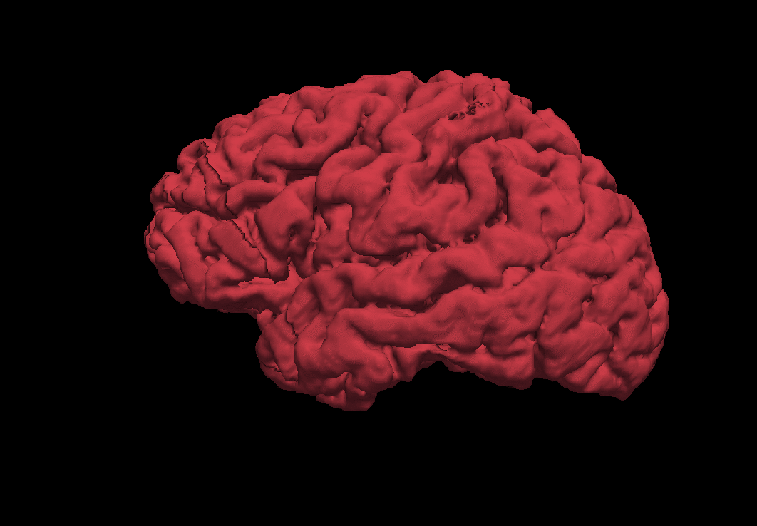
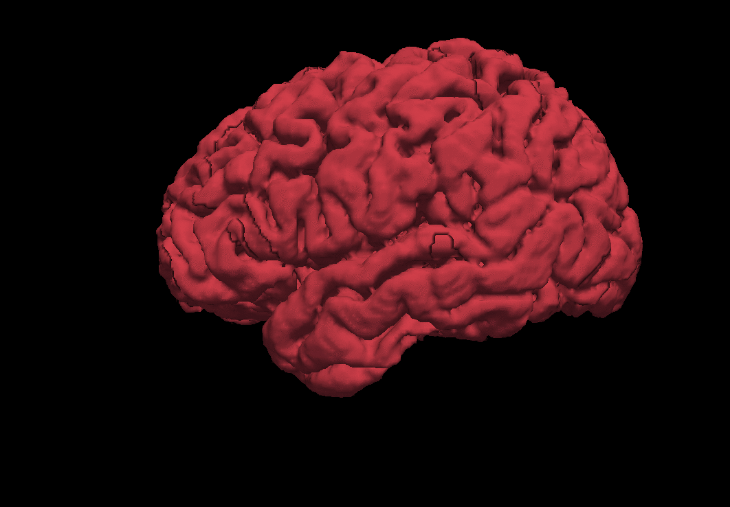
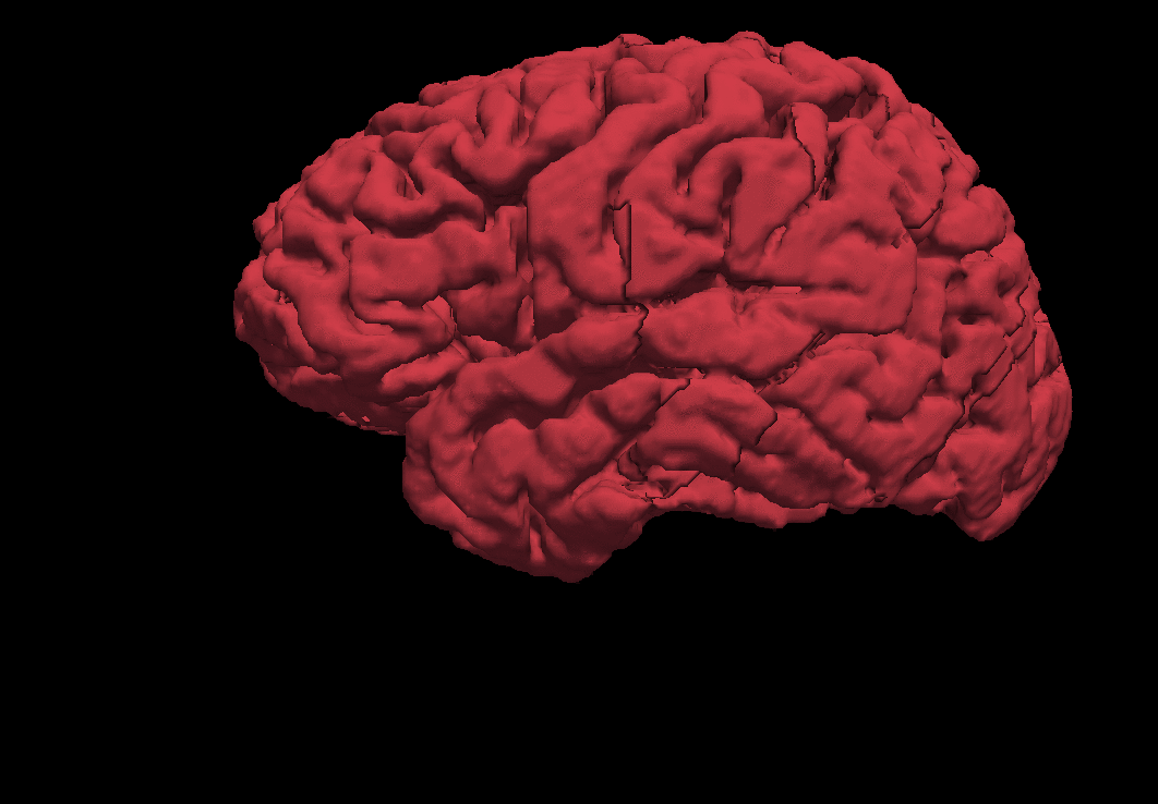
| sub-01 | sub-dd | sub-gg | sub-uu |
|---|---|---|---|
 |
 |
 |
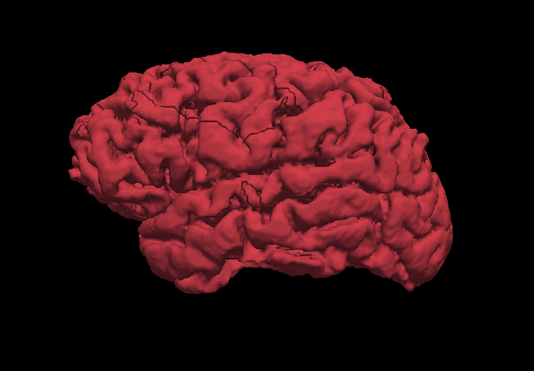 |
| $\begin{array}{cc}\text{lhCortexVol} & 234910.501 \ \text{rhCortexVol} & 236797.813 \ \end{array}$ | $\begin{array}{cc}\text{lhCortexVol} & 216503.982 \ \text{rhCortexVol} & 217683.821 \ \end{array}$ | $\begin{array}{cc}\text{lhCortexVol} & 240209.371 \ \text{rhCortexVol} & 243691.472 \ \end{array}$ | $\begin{array}{cc}\text{lhCortexVol} & 239790.613 \ \text{rhCortexVol} & 245022.333 \ \end{array}$ |
$\text{Table 1. 3D Visualization of Cerebral Cortex for Subjects}$
Conclusion
Each subject showed the different rate of change in the volume of the cerebral cortex. Although the left cortex volume is larger than the right cortex volume as following the common fact, the uniform rates were not observed. The project concluded that two major factors affects to this unmatched result; 1) unfamiliar movements to a subject can influence the omnidirectional brain regions 2) the process while collecting MRI data caused an error, resulting in hindering the precise measurement.
In summary, the paper reviewed the general idea of the motor cortex activation with the motor fMRI dataset. The analysis was conducted with the OpenNeuro dataset, and the results were visualized as 3D gif images with FreeSurfer.
References
\(\tag*{}\label{1} \text{[1] FreeSurfer. Accessed 18 Aug. 2024, https://surfer.nmr.mgh.harvard.edu/}\) \(\tag*{}\label{2} \text{[2] Veillette, J. and Nusbaum, H. (2024). motor-fmri. OpenNeuro. [Dataset] doi: doi:10.18112/openneuro.ds005239.v1.0.1}\) \(\tag*{}\label{3} \text{[3] Veillette, John P., Pedro Lopes, and Howard C. Nusbaum. "Temporal Dynamics of Brain Activity Predicting Sense of Agency over} \\ \text{Muscle Movements." Journal of Neuroscience 43.46 (2023): 7842-7852.}\)